1
Overview
Dynamic digital radiography is a new medical imaging modality that uses pulsed X-rays to acquire dynamic images of the chest and other regions. It adds temporal motion information to conventional plain radiography, thereby enabling functional assessment.
“DM-MODE,” one of the functions of Konica Minolta’s dynamic digital radiography analysis workstation, tracks the craniocaudal motion of the diaphragm and displays the displacement. The function is expected to be applied to patient management in the ICU (Intensive Care Unit), such as evaluation of diaphragmatic nerve palsy.1) 2) (Figs. 1, 2)
Fig. 1 Example of “DM-MODE”
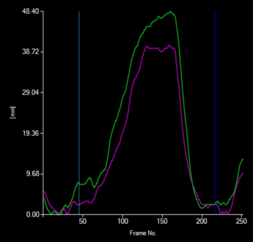
Fig. 2 Example of diaphragmatic displacement (Horizontal axis: Frame number, Vertical axis: Diaphragmatic displacement, Purple line: Right diaphragm, Green line: Left diaphragm)
Conventionally, the diaphragm position was detected using a rule-based method based on image contrast. However, detection accuracy decreased in images where the diaphragm boundary became unclear because of overlapping disease findings or cardiac silhouette enlargement. The artificial intelligence (AI)-based method introduced in this paper maintained high detection accuracy even for datasets that include images in which the diaphragm is not easily visible. This paper presents the configuration of DM-MODE, the issues in the conventional technology, and the improvements achieved using AI technology.
2
Details
■Configuration
The dynamic digital radiography system consisted of a pulsed X-ray generator and the dynamic digital radiography analysis workstation (KINOSIS), as shown below. Konica Minolta provides the AeroDR TX m01 as the pulsed X-ray generator (Figs. 3 and 4).
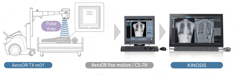
Fig. 3 Configuration of Dynamic Digital Radiography
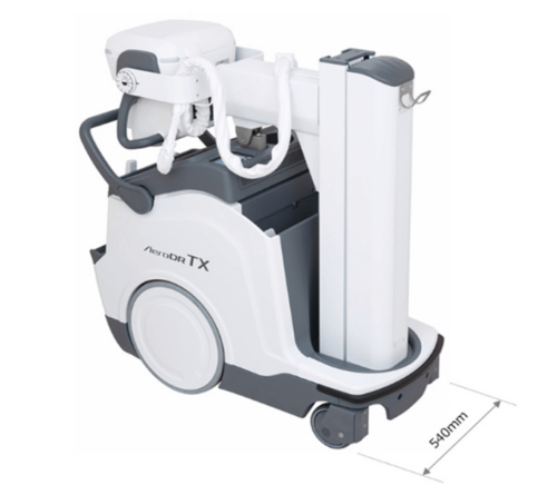
Fig 4. AeroDR TX m01
The dynamic digital radiography analysis workstation KINOSIS implements functions such as DM-MODE introduced in this paper, PH2-MODE for periodicity analysis synchronized with the heartbeat, and LM-MODE for displacement tracking within the lung fields (Fig. 5).
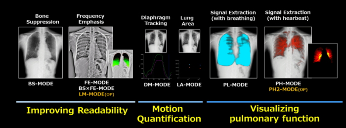
Fig 5. Analysis provided by KINOSIS (partial excerpt)
■Functions / Features / Applications
In DM-MODE, the conventional rule-based algorithm using edge information could misdetect the diaphragm position in images where contrast around the diaphragm is reduced because of overlapping disease findings or cardiac silhouette enlargement (Figs. 6(a) and 7(a)).
To address this issue, we trained an AI model using images selected and processed to represent diverse cases expected in clinical practice, based on interviews with multiple certified radiologic technologists. The model enables accurate localization of the diaphragm even in images with reduced contrast around the diaphragm. A convolutional neural network (CNN)–based model was adopted in this study. Because short processing time is critical in busy clinical settings, the model was optimized to be lightweight by reducing the number of parameters and the input image size.
Fig 6. Image with unclear diaphragm due to disease
Fig 7. Image with low contrast diaphragm due to enlarged heart
As a result of these efforts, the diaphragm identification success rate on the evaluation dataset improved from 80% to 95%, achieving a substantial increase in accuracy. In addition, processing time was reduced to approximately one-seventh to one-eighth of that of the conventional algorithm (Fig. 8).
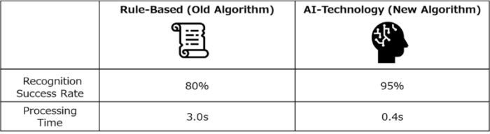
Fig 8. Evaluation results of recognition success rate and processing time
■Future outlook
AI-based diaphragm detection in DM-MODE achieved highly accurate recognition even in images with reduced contrast around the diaphragm.
The approach is expected to be particularly effective for severely ill patients and supine imaging cases, where contrast tends to decrease. For example, the method may contribute to expanding the clinical applications of X-ray video analysis in the ICU.