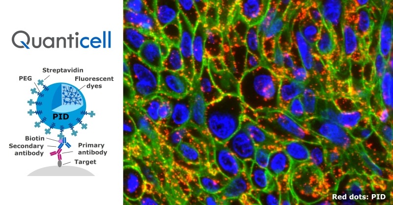
Quanticell: An immunostaining service enabling high-sensitivity, quantitative, and localization analysis
High-resolution imaging of target molecules using ultra high-bright fluorescent nanoparticles capable of single-particle detection
\Our expert team handles inquiries
on technical partnerships and joint research./
Value Proposition
An immunohistochemical staining and analysis service that achieves highly sensitive and highly reproducible quantitative fluorescence imaging, as well as high-resolution evaluation of localization/distribution
This analysis service provides a solution that visualizes and quantitatively measures specific molecules present in pathological tissue sections with high sensitivity and high reproducibility. It provides quantitative imaging that supports drug discovery by visualizing the distribution of proteins or candidate drug compounds and their effects on target tissues, cells, or molecular targets. Therefore, this service provides scientific evidence to elucidate mechanisms of action and evaluate the efficacy of candidate drug compounds.
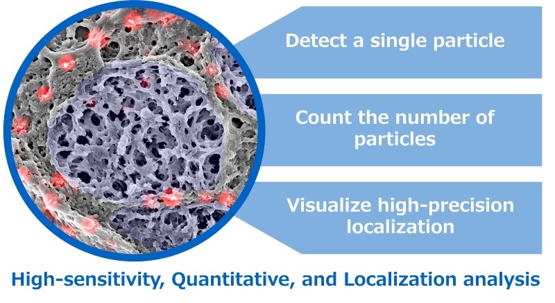
Key features of this solution include: (1) high-sensitivity detection that was difficult with conventional immunohistochemical staining, (2) highly reproducible quantitative analysis, and (3) the ability to image and analyze intratissue distribution and localization at high resolution.
(1) Konica Minolta developed fluorescent particles with high brightness and high photostability. Because a single particle can be detected, Quanticell can detect low-density molecules that were difficult to visualize using conventional staining techniques.
(2) Konica Minolta also developed an analysis algorithm that can count the “number of particles” as a physical and intuitive metric. Rather than simply converting fluorescence intensity into a numerical value, this algorithm enables stable acquisition of quantitative values corresponding to the amount of target molecules.
(3) Imaging with fluorescent particles provides high-resolution positional information and enables quantitative evaluation. Therefore, it is useful for quantitatively imaging intra-tissue distribution and higher-resolution localization analyses.
This solution provides information on particle counts and the particle density for each specimen, region, and cell.
Quanticell is offered as a service that supports advanced drug discovery research by combining advanced materials technologies from Konica Minolta, cultivated through prior development with proprietary analysis algorithms. If you are interested, please contact us for consultation using the inquiry form.
Technology Overview
This service comprises two main technologies.
The first technology is immunohistochemical staining. In this method, imaging is performed using an antigen–antibody reaction. Antibodies that specifically bind to target molecules are applied to a tissue section, and a fluorescent or chromogenic label is attached to the bound antibodies for visualizing the location of the target molecules. PID (Phosphor Integrated Dots) is used as a fluorescent label.
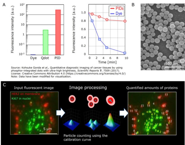
We have developed a technology that enables the stable production of PIDs with uniform particle size in which a large number of fluorescent dyes are densely encapsulated within a single particle. Compared with conventional fluorescent dyes, PIDs exhibit a more than 10,000-fold increase in fluorescence intensity with high stability.
Therefore, single-PID detection is possible, enabling the detection of low-density molecules that are difficult to detect with conventional methods.
The second technology is a quantitative analysis algorithm. Our proprietary algorithm automatically counts the number of particles in PID-stained images, enabling highly reproducible quantification of target molecules. Further, because the particles appear as dot-like bright spots, high-resolution localization/distribution analysis is possible.
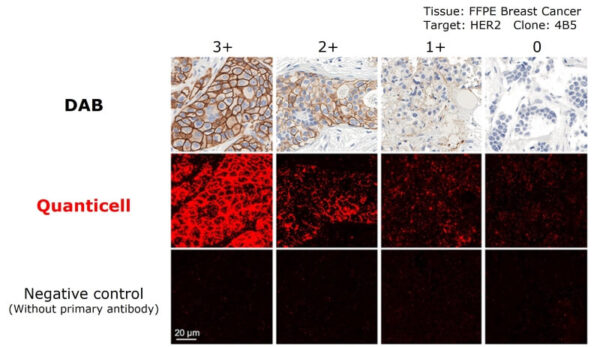
Use case 1: High-sensitivity quantification (comparison with DAB staining)
The first use case is high-sensitivity quantification in immunohistochemical staining.
Immunohistochemistry is an imaging technique based on antigen–antibody reactions. Generally, DAB staining is widely used, wherein target molecules are visualized in brown. For example, in breast cancer, the HER2 protein expressed in tumor tissue is stained using DAB, and the HER2 score (0, 1+, 2+, or 3+)—based on staining intensity and the proportion of positive cells—is evaluated to guide treatment decisions. However, DAB staining has limitations in sensitivity and quantification. A key challenge of evaluation with DAB staining is that it relies on visual assessment by pathologists, which makes the results prone to inter-observer variability.
In contrast, Quanticell uses fluorescent nanoparticles (PID) to detect target molecules with higher sensitivity. Furthermore, because the analysis is based on counting particles, it enables quantitative evaluation with high reproducibility, even for low-abundance targets.
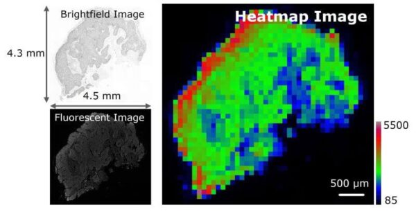
Use case 2: Quantitative fluorescence imaging (macroscopic distribution analysis of the entire tissue section)
The second use case is quantitative analysis of whole-tissue fluorescence images. intratissue distribution analysis using fluorescence imaging.
Quanticell can capture fluorescence images of the entire tissue section and quantify the macroscopic distribution of target molecules across the section. For quantitative visualization, PID counts can be represented as pseudo-color heat maps. This approach enables quantitative analysis of protein abundance and distribution on the tissue section, as well as assessment of whether candidate drug compounds have reached their target regions.
Use case 3: Quantitative localization imaging (microscopic distribution analysis at the organelle level)
The third use case is quantitative analysis of localization/distribution using high-resolution imaging.
Because PID appears as dot-like bright spots, it can provide high-resolution positional information. By combining confocal microscopy, Quanticell enables quantitative evaluation of localization at the cellular and organelle levels. As an example, the figure below shows an analysis of the intranuclear and extranuclear distribution of Estrogen Receptor (ER) stained with PID. This method can be applied in drug development to elucidate mechanisms of action, including the intracellular localization of administered drug candidate compounds.
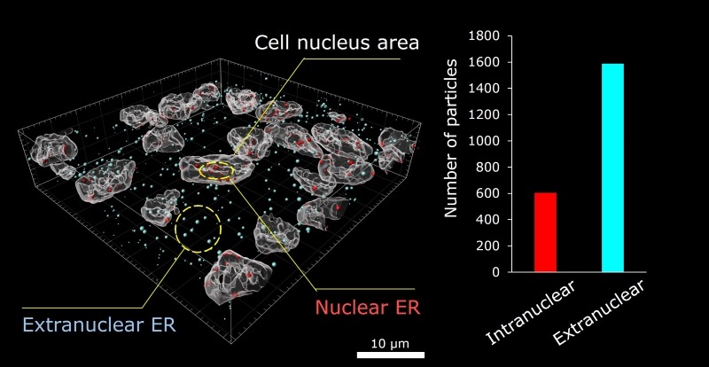
3D confocal fluorescence image (left) and digitally analyzed image (right)
Left: white area, cell nucleus; red, PID
Right: red dots, intranuclear ER; light blue dots, extranuclear ER
Use case 4: Simultaneous quantification (simultaneous detection of target molecules using two-color PID)
The fourth use case is the simultaneous quantification of target molecules stained with two-color PID.
Quanticell can simultaneously stain and detect multiple targets using red and green PID, enabling quantitative comparison of two targets within the same tissue section. For example, by differentially labeling a drug candidate compound and its therapeutic target protein using two-color PID staining, this method can be applied to pharmacological efficacy assessment.
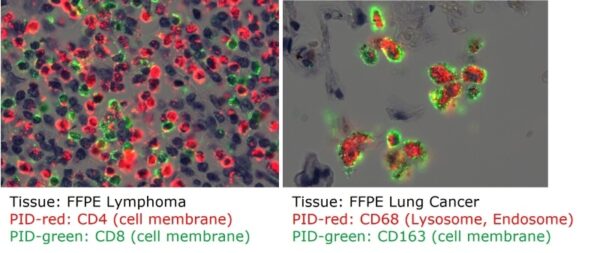
Further, multiplex staining of two or more targets can be achieved up to five fluorescence colors by combining PID with general fluorescent dyes. This enables quantitative analysis of PID-labeled target molecules distributed within specific cells or subcellular organelles that have been stained with fluorescent dyes.
Application areas and keywords
Application areas: Drug discovery
• Pharmacokinetics
• Efficacy/pharmacology
• Toxicity/safety
• Biomarker exploration, etc.
Keywords
• Patient stratification (biomarkers)
• Correlation with pharmacological efficacy (biomarkers, drug candidate compounds)
• In vivo drug distribution (drug candidate compounds)
• Mechanism-of-action (MoA) elucidation (drug candidate compounds)
Other potential applications
• Cell-based analysis
*Quanticell is a registered trademark of Konica Minolta, Inc.
For more technical information
- KONICA MINOLTA Technol. Rep. 2021, 18, "Fluorescent nanoparticle PID (Phosphor Integrated Dots)"
- KONICA MINOLTA Technol. Rep. 2017, 14 "HSTT (High Sensitive Tissue Testing)"
- News release on issuance of an international standard
- NEDO "Practical Realization Document" interview article
- Reference 1: Scientific Reports 7, 7509 (2017)
\Our expert team handles inquiries
on technical partnerships and joint research./