1
Overview
MIMPS stands for Medical Image Management and Processing Systems, and it is a system that automates the management and processing of medical images to reduce the workload of radiologists and enhance the efficiency of the image diagnostic workflow.1) One example of MIMPS utilizing AI technology (hereafter referred to as MIMPS-AI) is the Synchronization Assist feature included in our PACS (Picture Archiving and Communication System) product, the medical image management system FINO.VITA.GX. This feature automatically synchronizes and displays the anatomical positions of current and past CT examination images, making it easier to confirm temporal changes (Fig. 1). Additionally, we are advancing the development of the Reading Report Analysis Support technology as a means to enhance medical safety and efficiency. This technology aims to structure key information such as findings, diagnoses, and locations from plain text recorded in reading reports, enabling efficient comprehension of findings and appropriate management of reports with noted findings. This report introduces the above two functions and their features.
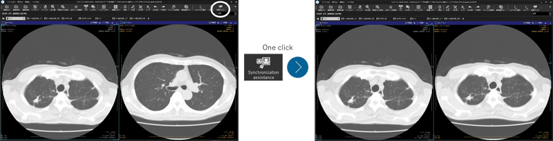
Fig. 1 Comparison of images by synchronization assistance
2
Details
■Configuration
1. Synchronous Assist
The CT image synchronous assist function consists of organ shape analysis processing and display processing. The analysis processing is executed at the timing when the image is received by PACS, analyzing the CT image (on a series basis) to extract information (landmarks) related to organ shapes. Subsequently, when the image is displayed, display control is performed based on this analysis result in the PACS viewer, enabling efficient diagnosis.
2. Report Structuring
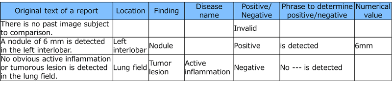
Report structuring is the process of systematically organizing the areas, findings, disease names, affirmative or negative judgments, and numerical information described in chest CT image reports. This process involves analyzing the findings and diagnostic sentences on a sentence-by-sentence basis. Specifically, it performs morphological analysis and phrase identification based on a dictionary, identifies disease names, findings, and areas, makes affirmative or negative judgments (determination of findings), and extracts structured information.
■Functions / Features / Applications
1. Synchronization Assist
The function that recognizes landmark positions from CT images is a feature that extracts specific anatomical points using three-dimensional image analysis technology that utilizes Deep Learning. Based on these landmark positions, alignment can be performed on the active image, allowing for comparison of images at the same position. The alignment operation can be completed with a single click, which is expected to provide rapid and efficient diagnosis.
2. Report Structuring
The proposed report analysis method provides a function to efficiently structure the information from chest CT image reports. Notably, it achieves high structuring accuracy along with rapid processing. The accuracy of findings and disease name determinations for indicated observations is 97.3%, and the processing time per report on a general notebook PC with a CPU equivalent to Intel Core i5-8250U is achieved in less than 0.5 seconds (Table 1). Since analysis results can be obtained in real-time, efficient understanding of findings in practical operations and appropriate management of reports with findings are expected.
Table 1 Example of report structuring
■Future outlook
Radiology operations using MIMPS-AI are expected to increase efficiency in various aspects and help reduce the workload of physicians by strengthening the synchronization assistance function through the expansion of parts covered by MIMPS-AI and by improving the accuracy and upgrading the function of the report analysis solution.