1
Overview
Chest X-ray imaging is a common examination widely practiced around the world and is known for its high volume of radiographs. However, radiogram interpretation is challenging due to the diverse range of target diseases, making it one of the fields where computer-assisted diagnosis can be particularly effective. CXR Finding-i, an AI-assisted diagnostic support system for chest X-ray images, was released in November 2021 as a solution to help prevent physicians from overlooking findings and to support clinical practice. Subsequently, an improved version with enhanced accuracy was released in October 2024. As of December 2024, the system has been deployed at more than 700 medical institutions across Japan. Utilizing Deep Learning, an AI technology, it extracts characteristic patterns from chest X-ray images and marks areas where target findings are suspected. This paper presents an overview of the developed AI technology, results from interpretation experiments conducted with 12 physicians, and the system configuration.
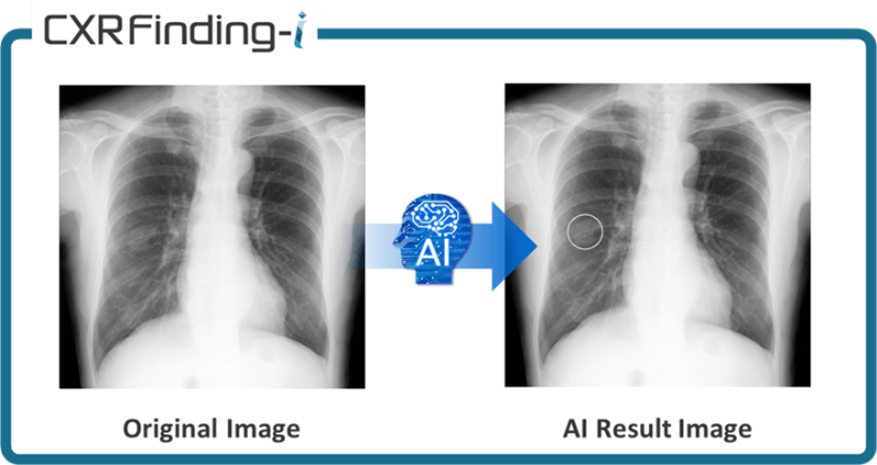
Fig. 1 Overview of CXR Finding-i
2
Details
■Configuration
CXR Finding-i (hereafter referred to as “this product”) is software as a medical device designed to be installed in various Konica Minolta systems (Fig. 2). Through these systems, this product performs AI analysis on frontal chest X-ray images received from X-ray imaging devices. The system then sends the analysis results to picture archiving and communication system (PACS) and other devices.
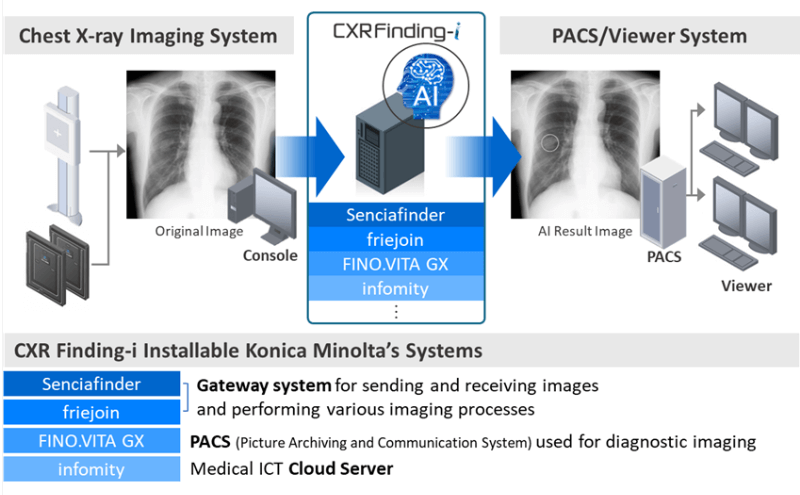
Fig. 2 System Architecture and Integration Overview of CXR Finding-i
There are many technologies for detecting suspected abnormal findings in medical images. However, this product incorporates not only deep learning technology but also Konica Minolta’s proprietary image processing technology and expertise in medical imaging into its algorithms (Fig. 3). The deep learning model architecture is designed to achieve a balanced extraction of both global and local features of abnormal findings, while also optimizing analysis processing time and memory usage. Recognizing that data is a critical element in improving deep learning model performance, we placed significant emphasis on data collection during product development. By collecting and utilizing hundreds of thousands of high-quality images from major hospitals and health screening facilities both within Japan and internationally for training, we have substantially enhanced the model’s robustness.
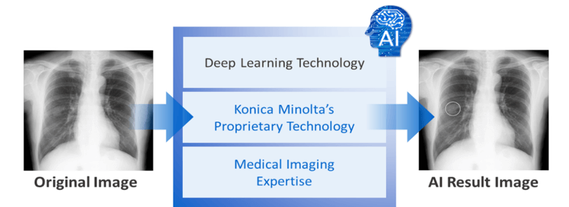
Fig. 3 AI Components Powering CXR Finding-i
As a result of these efforts, the improved version released in October 2024 achieved detection sensitivity of 84% for nodules and masses and 85% for consolidations. The specificity reached 88%, a 19-point improvement from the 69% of the initial version. In an observer study involving 12 physicians, the Figure of Merit (FOM), which measures diagnostic accuracy, showed a statistically improvement from 0.756 to 0.878 when using this product. Additionally, the physicians’ average sensitivity increased by 14 percentage points, from approximately 75% without the product to approximately 89% with it. These results demonstrate that the use of this product statistically significantly improves physicians’ radiographic interpretation performance (Fig. 4).
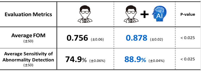
Fig. 4 Reader Study Results
■Function / Features / Applications
This product is compatible with images captured by other manufacturers’ systems and can accept them as input images. The analysis results are presented as simple circular marks overlaid on the original images, attracting physicians’ attention to areas of interest and serving as reference during interpretation (Fig. 5).
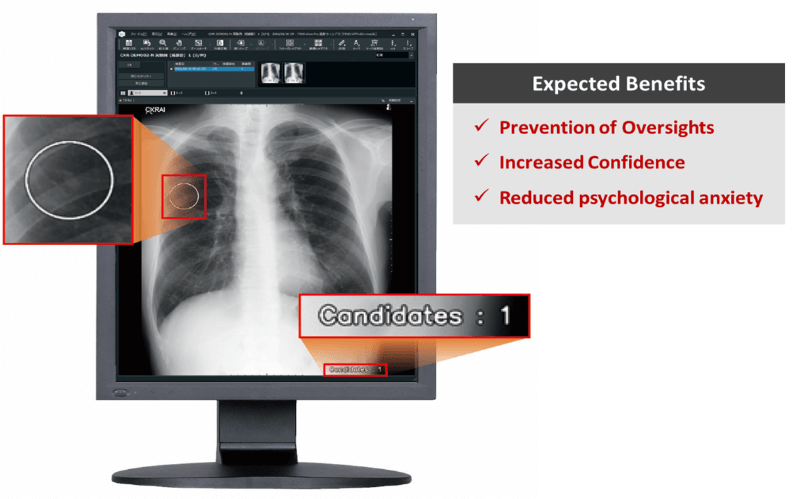
Fig. 5 AI Result Image Sample
Additionally, this product can be integrated with FINO.Report in the FINO.VITA.GX system. This integration enables the system to compare physicians’ interpretation reports with AI analysis results, check for discrepancies in findings, and notify users (Fig. 6). This functionality provides opportunities for physicians to identify potential oversights in their findings, thereby contributing to further improvement in healthcare quality.
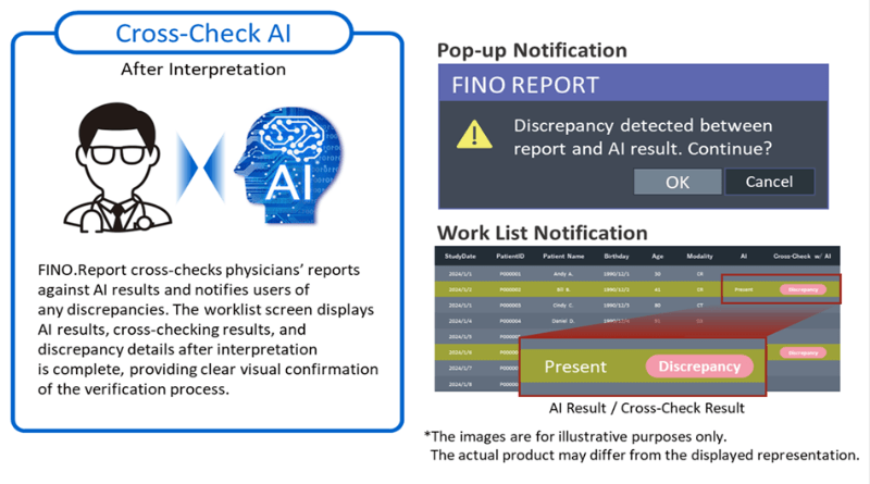
Fig. 6 Integration of CXR Finding-i with FINO.Report: Cross-checking System
■Future outlook
Konica Minolta has developed image processing technologies to support physicians in interpreting chest X-ray images, such as Bone Suppression and Temporal Subtraction. Moving forward, we plan to actively incorporate cutting-edge technologies including generative AI and foundation models, building upon our proprietary technologies to enhance products that address diverse needs in clinical settings, such as reducing physician workload and improving operational efficiency.