1
Overview
Diagnostic ultrasound systems are used across a wide range of clinical departments because they enable noninvasive, real-time observation. One application in orthopedics involves determining surgical indications for diseases such as carpal tunnel syndrome and cubital tunnel syndrome, in which nerve cross-sectional area is used as one of the assessment indices1). Conventionally, measuring nerve cross-sectional area on a diagnostic ultrasound system required physicians to manually trace the contour of the nerve region, which was time-consuming and labor-intensive.
To address this issue, we developed the “Nerve Cross-Sectional Area Measurement” function, which uses artificial intelligence (AI) to identify nerve regions in ultrasound images and semi-automatically help measure nerve cross-sectional area. This function has been implemented in the diagnostic ultrasound system SONIMAGE UX1. It reduces operational burden and helps improve the examination workflow of physicians.
2
Details
■Configuration
This function comprises a sequence of processes that identify the nerve region in an ultrasound image and semi-automatically measure its cross-sectional area. The system operates through a processing flow comprising AI-based detection of the nerve region, selection of the target nerve for measurement, and calculation of the measured value (Fig. 1).

Fig. 1 System overview of this function
■Functions / Features / Applications
The key features of this function are automatic recognition of the nerve region and semi-automated measurement of cross-sectional area. This function is intended for diagnostic situations where quantitative evaluation of nerve cross-sectional area is required, such as assessment of the median nerve in carpal tunnel syndrome. With conventional manual tracing, physicians must draw a line following the contour of the nerve region, and the measurement task is time-consuming (Fig. 2a). In addition, if the contour of the nerve region requires correction, the physician must perform the manual tracing operation again.
In contrast, this function enables the physician to simply freeze the image and press the measurement button. The AI recognizes the nerve region and calculates the cross-sectional area (Fig. 2b).
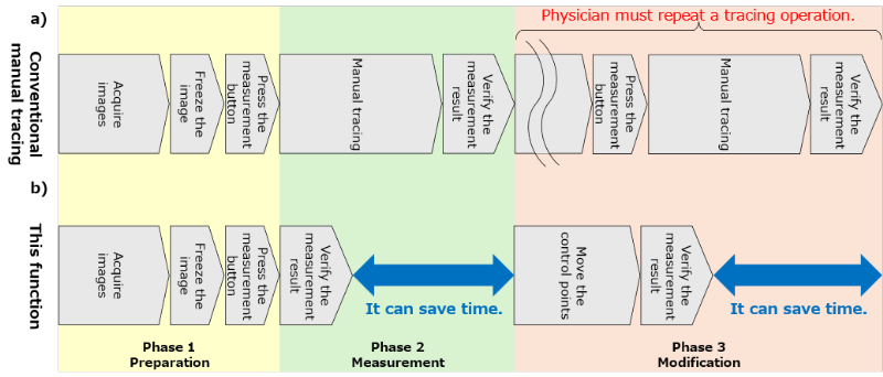
Fig.2 (a) Operation flow of conventional manual tracing and (b) operation flow using this function for nerve cross-sectional area measurement
The measurement results are displayed on the screen as the contour of the nerve region and the numerical value of the cross-sectional area, allowing the physician to review them (Fig. 3). After verifying the accuracy of the measurement result, the physician can easily fine-tune the contour by moving the control points displayed on the screen (the crosshair points in the right panel of Fig. 3) via touch operation, as needed. The spacing between control points was designed to be wider than a fingertip to reduce stress during touch operation.
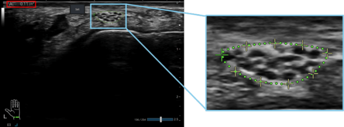
Fig.3 Example display of the automatically measured nerve contour (green dashed line) and the cross-sectional area value (inside the red box)
Thus, the function employs a semi-automated measurement approach that balances automation with physician control while prioritizing usability during manual adjustment. This design reduces measurement time and physician workload. In addition, when multiple nerve regions are detected, the system automatically selects the most appropriate region according to criteria consistent with clinical practice. This allows efficient measurement of the nerve region that the physician intends to evaluate based on probe positioning.
■Future outlook
This function utilizes AI-based nerve recognition technology as a practical measurement tool for clinical use. It addresses clinical needs for quantitative diagnosis and contributes to improving examination efficiency. At present, the function is limited to use after image freezing; however, future development aims to enable real-time application during examinations. We will continue to advance diagnostic ultrasound systems through the development of functions that further reduce the workload of physicians.