Dynamic digital radiography and image analysis technologies
A new X-ray imaging method that visualizes and quantifies organ movements
\Our expert team handles inquiries
on technical partnerships and joint research./
A breakthrough in chest radiography through visualization of movement
Without being bound by the conventional common sense that static imaging should be used for plain radiography for a radiographic diagnosis, we contribute to the diagnosis of lung diseases, which pose a serious problem in Japan and other countries, such as chronic obstructive pulmonary disease (COPD), pneumonia, and lung cancer by providing new information of movement. We are developing visual information on respiratory movements of the diaphragm, ribs, and other structures in the lung region as well as functional imaging of ventilation, blood circulation, and other functions using our original image processing technology. This enables the acquisition of more information than that obtained with static imaging, thus accelerating the elucidation of various pathologies, start of treatment, and assessment before and after therapy, and contributing to more efficient medical care and the reduction of medical costs.
Technology Overview
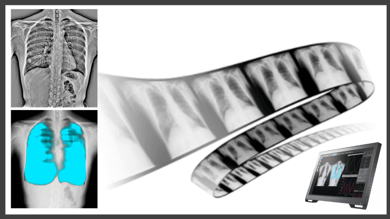
We deliver the new value of dynamic radiography with its new technologies. The Dynamic Digital Radiography system is a completely new system that comprises a dynamic X-ray image analysis workstation, a portable digital radiography (DR) unit, and a typical digital X-ray device, which is also used for conventional plain radiography. This system applies serial X-ray pulses to the body and displays a series of frame-by-frame images to create a dynamic image. We are developing our own image processing technology for X-ray movie analysis.
Dynamic X-ray image processing technology
Research and development of visualization and quantification of ventilation and blood circulation distribution in the lung region
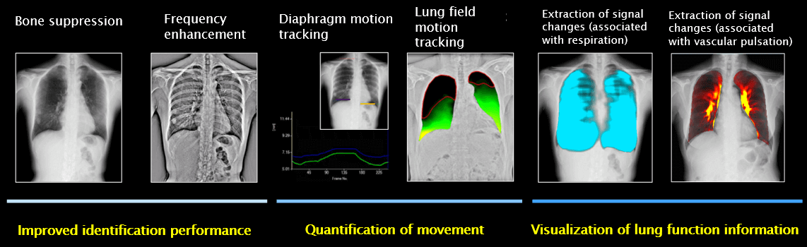
Fig. 1: Group of images for digital dynamic radiography
(1) Research and development of visualization and quantification of movement
We apply static imaging processing technology to dynamic imaging to improve clarity and quantify movements, and thus provide various image groups and obtain information that cannot be obtained from static images to infer symptoms and assess function. Fig. 1
(2) Research and development of visualization of biological functions
• Basic principle: The X-ray transmission per unit volume of the lung alveoli and blood vessels changes with respiration and heartbeat. This process measures density changes of structures in the lung region and may be useful in assessing lung function. Fig. 2
• Visualization of signal change (associated with vascular pulsation)
This process visualizes signal changes in the lung region in synchronization with the heartbeat during the following procedures: Fig. 3
1. Identifying the lung and the heart regions.
2. Determining the time frequency of signals in the left ventricular region.
3. Identifying the maximum diastolic frame in the heart region.
4. Extracting only the signals of the time frequency determined in 2 from signals in the lung region.
5. Subtracting the signal in the maximum diastolic frame identified in 3 from the signal of each frame in the lung region.
[Key technologies] Lung and heart recognition processing, time filtering processing, and reference frame subtraction processing.
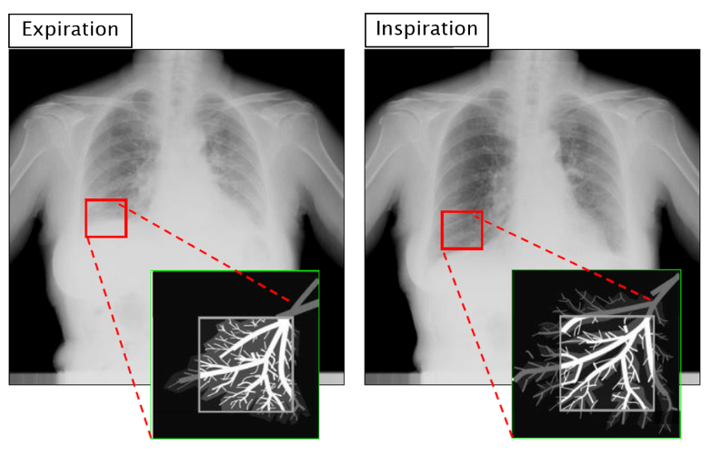
Fig. 2: Visualization of biological functions; the basic principle of ventilatory function
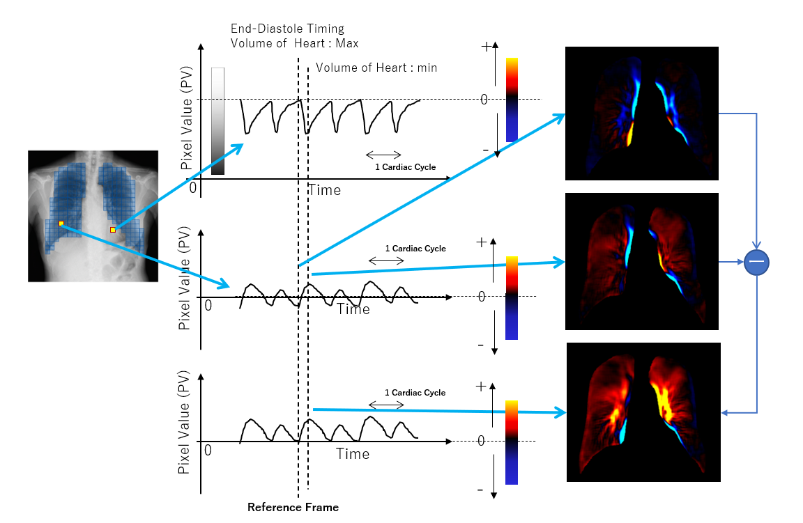
Fig. 3: Visualization of biological functions; an example of a signal change (associated with vascular pulsation) analysis algorithm.
\Our expert team handles inquiries
on technical partnerships and joint research./