High-sensitivity quantitative protein analysis technology
Target proteins visualized by proprietary fluorescent particles
\Our expert team handles inquiries
on technical partnerships and joint research./
A new method of tissue staining for quantitative and highly sensitive analysis
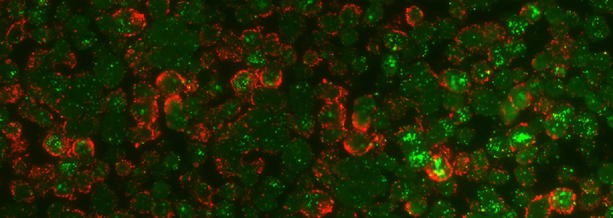
Using Konica Minolta’s proprietary high-intensity fluorescent particles, this technology sensitively detects a target protein, which is expressed in and outside cells in slices of human or animal pathological samples, or drug bound with the target protein. It enables the target protein to be quantified with high sensitivity, overcoming difficulties faced by conventional technologies. The technology also provides supplemental information to complement scientific evidence for drugs by means of localized analysis leveraging detailed image analysis.
Technology Overview
The features of PIDs include a uniform particle size, higher intensity than fluorescent pigments or quantum dots, and superb stability when irradiated by an excitation light source. Thanks to these features, this technology enables quantitative, high-sensitivity analysis not possible with conventional methods. Regarding immunostaining techniques, it follows a similar process to conventional methods, staining in order of the primary antibody, the secondary antibody, and PIDs. Accordingly, it requires no special reagent or antibody. Bright-field and fluorescent images obtained from stained specimens are analyzed by Konica Minolta’s proprietary software to enable quantitative evaluation. Bright-field images are used to define the nuclear region and measure the number and area of cells through digital processing based on a nuclear recognition algorithm. Fluorescent images are used to calculate the amount of the target protein based on the fluorescence intensity of bright points.
\Our expert team handles inquiries
on technical partnerships and joint research./