Medical ultrasound imaging technology
Providing all-in-one style from consultation to treatment using ultrasound diagnosis
\Our expert team handles inquiries
on technical partnerships and joint research./
A compact and high-quality-imaging medical ultrasound
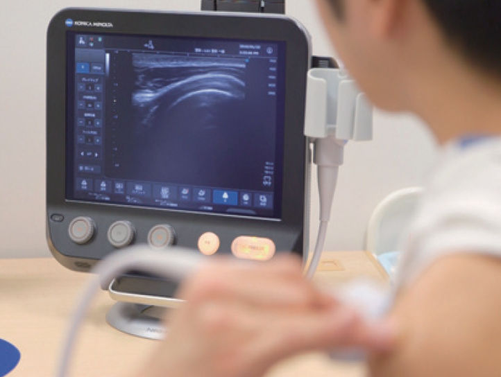
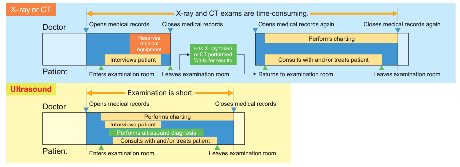
It may seem as if diagnostic ultrasound imaging is equivalent to abdominal examination and fetal monitoring. Since ultrasound systems can also observe movement easily and in real time, they are increasingly being used in the field of musculoskeletal and sports medicine. If ultrasound devices that can provide clearer images are used, it is possible to grasp the condition of musculoskeletal structures in patients with painful shoulders, low back, or joints in more detail. Once the cause of pain is identified, the physician can treat it by injecting a drug solution into the affected site while monitoring the ultrasound image in the outpatient consultation room. Patients can save waiting time for many examinations and receive treatment as soon as the cause of the pain has been established, rather than having to undergo follow-up observation with patches and pain relievers. Konica Minolta’s diagnostic ultrasound system can contribute to the evolution of such new types of outpatient care.
Technology Overview
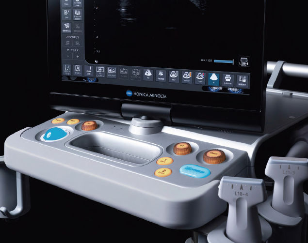
By combining a material development technology that can control the acoustic physical properties of a material as desired by accurate manufacturing, we have successfully developed a wideband, high-sensitivity, high-frequency transducer with unprecedented wideband properties. To leverage the transducer features, we have produced a high-quality imaging engine by integrating our original ultrasound transmitting/receiving technology and our image processing technology. The engine and intuitive user interface are housed in a compact device. By downsizing the device, we have achieved a light-weight, power-saving device. Due to its high operability with the minimum number of buttons and a touch panel, our diagnostic ultrasound system can be provided for various situations and users.
Acoustic and material design technologies for the high-sensitivity wideband transducer
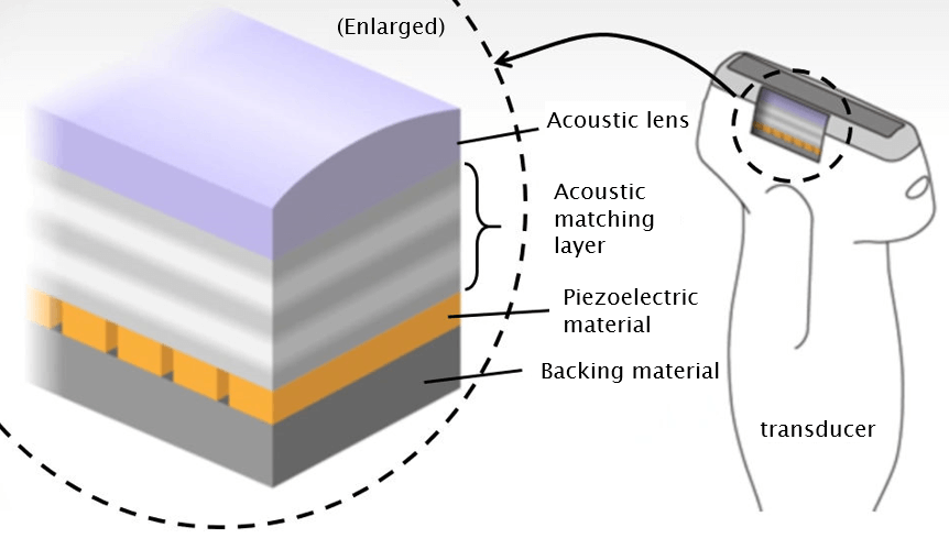
The transducer not only transmits but also receives ultrasound waves. To efficiently send ultrasound signals, which are based on vibrations created by applying a voltage to a piezoelectric material, into the body and produce an image of the signals from deep within the body, we have meticulously designed the acoustic materials of the transducer as well as the signal waveforms to be transmitted. Our technologies have overcome existing electrical, acoustic, and structural problems and ensure both the high sensitivity and wideband performance of the transducer.
High-quality signal transmitting/receiving technology
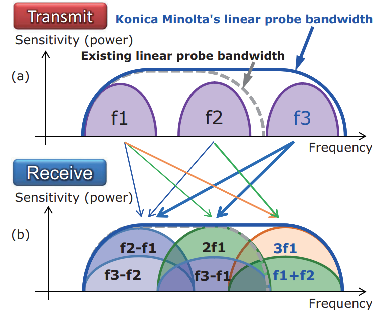
We are developed high-quality signal transmitting/receiving technology that transmits three independent frequency components simultaneously and receives multiple harmonic components. Making maximum use of the ultra-wideband properties of the transducer, this technology can transmit and receive frequency components on the high frequency side, which has been impossible with conventional methods. This allows us to use various signals entering the frequency band, such as difference and sum tones and harmonics, to generate images with top-level resolution. We have also improved the S/N ratio in the superficial area although the device is portable.
Realizes the production of a wide range of high-quality images from the superficial to the deep layers of the body
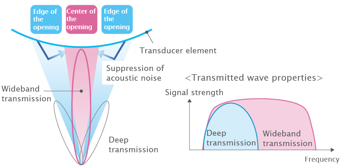
Diagnostic ultrasound devices determine transmission conditions such as transmission time interval, focal depth, and transmission opening size depending on the display depth, focus point, and other set values. Under these conditions, an optimal voltage determined taking human safety into account is applied to the piezoelectric element. To form a transmission focus in a deep layer, ultrasound waves are usually transmitted through a relatively large aperture. At a superficial depth, transmitted waves passing through the outside of the receiver focus area produce reflected and scattered waves, partly responsible for acoustic noise generation in the superficial area of the image. Excessive acoustic output outside the receiver focus area, which is not involved in image creation, generates unnecessary heat, resulting in limited transmission voltage. With this technology, ultrasound waves are simultaneously transmitted at different frequencies for superficial and deep layers, thus providing higher-quality images.
Improved needle visibility for injections
Ultrasound images can be displayed in real time, so the physician can inject a drug solution into the target area or collect tissue samples while monitoring living tissues and can also operate various devices while using ultrasound images as a guide. These are called ultrasound-guided procedures. Inserting a needle into the body involves the risk of damaging non-targeted blood vessels or nerves, which must be avoided. We are thus developing technology that recognizes special scattered waves from the needle tip or needle-like objects that move in a straight line by processing of the image and enhancing of these images. Not only when a needle is inserted parallel with the imaged plane, but also in a perpendicular direction, can this technology provide a clear image of the moment when the inserted needle crosses the slice plane, thereby supporting more safe procedures.
\Our expert team handles inquiries
on technical partnerships and joint research./